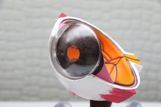
Visual defects lead to various kinds of retinal diseases, which are considered the major causes of low vision.
Ophthalmology is a medical specialty that deals with the eye, visual defects, and ocular diseases that may be related to it.
According to JLR eye hospital experts visual defects are visual disturbances that will prevent a person from seeing an object in focus. These defects can be corrected by wearing glasses or laser operations.
Ocular pathologies are more numerous and more diverse. Prevention, detection, and treatment or surgery of these are essential for the health of your eyes.
Visual Defects
Myopia
In Myopia, the image of the observed object is formed in front of the retina, so it is seen blurred.
Myopia affects distance vision but can also interfere with near vision if it exceeds a certain power. Correction is done with concave lenses.
The prevalence of myopia in the population is 15 to 20% in Western countries, however, its frequency seems to be increasing worldwide and particularly in Asia.
Hyperopia
Hyperopia prevents near vision (reading, telephone, etc.) but generally hinders distance vision. The correction is done with convex lenses.
Astigmatism
Astigmatism is characterized by an irregular shape of the cornea resulting in the focusing of light images at two different points of the eye, which therefore implies a deformation of the image. A round dot will be seen as a line by a person with astigmatism. This ametropia affects both far and near vision.
Correction is done with toric lenses.
Presbyopia
Presbyopia is a natural process of aging of the lens characterized by the progressive loss of the eye’s power of accommodation.
It results in the reduction of the elasticity of the lens as it ages. The latter is less able to modify its curvature. The image of nearby objects is therefore formed behind the retina. Correction is done with convex lenses in VP.
Glaucoma
Glaucoma is an optic neuropathy characterized by a rise in eye pressure causing damage to the optic nerve. More simply, it is an ocular pathology that will damage the optic nerve, the nerve allowing the transfer of visual information to the brain, its alteration by excessive eye pressure will cause damage to the visual field.
Glaucoma is a blinding disease if it is not detected and treated in time. Its insidious evolution without apparent symptoms or functional discomfort felt by the patient requires regular consultation with an ophthalmologist from the age of 40 in order to detect any signs of appearance.
Glaucoma is too much pressure inside the eye. This pressure can damage the optic nerve, the nerve that transmits information between the eye and the brain. Untreated optic nerve damage can lead to loss of vision and can even cause blindness.
Pressure in the eye may increase due to iridocyclitis or corticosteroids used to treat iridocyclitis. Checking for glaucoma should be part of a child’s regular eye exams by an ophthalmologist or optometrist.
Since glaucoma can develop in those with iridocyclitis, prevention is very important! If a child has glaucoma, they should be treated by an eye doctor. Treatment begins first with eye drops or medication taken by mouth.
Glaucoma does not reduce (except very late) central visual acuity, but it does reduce your ability to see to the side, little by little holes appear in your field of vision.
In the case of glaucoma, different treatments are possible (hypotonic eye drops, laser, or surgical treatment) generally allowing to limit or stop its evolution.
Regular monitoring of ocular fundus pressure and treatment, as well as additional examinations, are nevertheless always necessary.
AMD
AMD stands for Age-Related Macular Degeneration, which means “too” rapid aging of the macula (central part of the retina and therefore of vision). Tobacco smoke is a risk factor for AMD. AMD can lead to loss of central vision while leaving peripheral vision intact.
The atrophic form of AMD evolves slowly towards a decrease in visual acuity. It is characterized by the progressive disappearance of the cells of the pigmentary epithelium of the retina.
Early detection allows better follow-up and treatment
Diabetic retinopathy
Diabetic retinopathy is one of the leading causes of blindness worldwide in adults.
It is the result of retinal vascular disorders in the diabetic person whose disease has been unbalanced for a long time.
The early stages are characterized by retinal vascular occlusions and dilatations. Then it progresses to proliferative retinopathy with the appearance of new blood vessels in the retina.
A balancing of diabetes is important to limit the evolution of retinopathy. In the case of diabetes, regular follow-up with the ophthalmologist is necessary to check for any possible sign of the appearance of retinopathy.
If diabetic retinopathy is detected, it is important to regularly monitor the evolution with an ophthalmologist.
Cataracts
Cataracts can also be a side effect of certain medications used to treat JIA, such as corticosteroids. These side effects depend on the dose of corticosteroid and how long this medication is taken. Some children and adolescents seem to be more vulnerable than others, for unknown reasons.
Although steroid eye drops used to treat iridocyclitis can cause cataracts, the risk of developing cataracts when you have iridocyclitis is higher than if you did not use these drops.
Treatment for cataracts includes surgery to remove the cloudy lens from the eye. It is a day surgery, which means that the child or teenager does not need to spend the night in the hospital.
There are no medications or laser treatments for cataracts, and cataracts cannot simply go away. However, in some cases of less severe cataracts, when vision is not affected, no treatment is needed.
What examinations for screening?
The diagnosis of these diseases is carried out by the ophthalmologist. Several techniques can be used, in addition to the simple measurement of visual acuity:
- The fundus, a simple examination allowing direct observation of the retina
- Tomography of the eye by optical coherence scanner (or OCT), a true scanner allowing to obtain a cross-sectional view of the retina
- Angiography, a complimentary examination to observe the vessels of the retina, is important to detect possible diabetic retinopathy
In case of suspicion of genetic retinal disease, one can resort to tests in the case of hereditary retinopathies in order to determine which gene is altered in the pathology.


